
Science & Tech
Epic science inside a cubic millimeter of brain
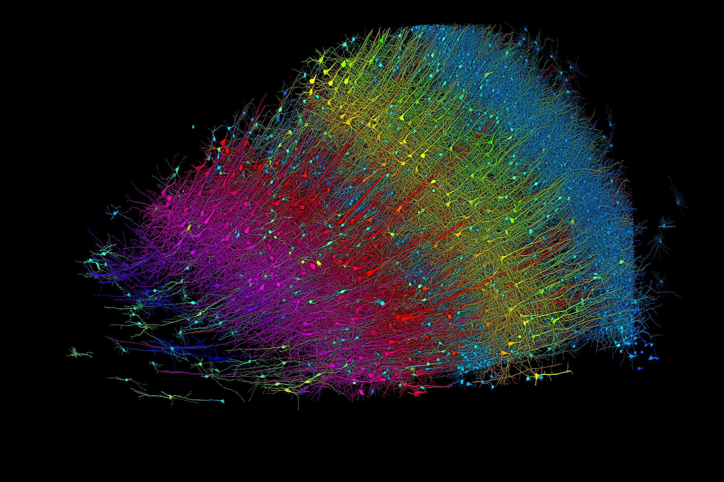
Six layers of excitatory neurons color-coded by depth.
Credit: Google Research and Lichtman Lab
Researchers publish largest-ever dataset of neural connections
A cubic millimeter of brain tissue may not sound like much. But considering that that tiny square contains 57,000 cells, 230 millimeters of blood vessels, and 150 million synapses, all amounting to 1,400 terabytes of data, Harvard and Google researchers have just accomplished something stupendous.
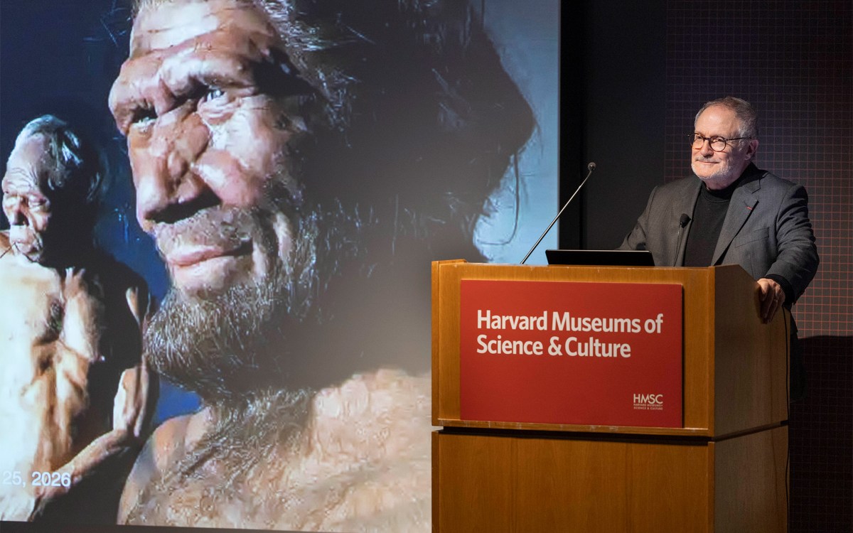
Led by Jeff Lichtman, the Jeremy R. Knowles Professor of Molecular and Cellular Biology and newly appointed dean of science, the Harvard team helped create the largest 3D brain reconstruction to date, showing in vivid detail each cell and its web of connections in a piece of temporal cortex about half the size of a rice grain.
Published in Science, the study is the latest development in a nearly 10-year collaboration with scientists at Google Research, combining Lichtman’s electron microscopy imaging with AI algorithms to color-code and reconstruct the extremely complex wiring of mammal brains. The paper’s three first co-authors are former Harvard postdoc Alexander Shapson-Coe, Michał Januszewski of Google Research, and Harvard postdoc Daniel Berger.
The ultimate goal, supported by the National Institutes of Health BRAIN Initiative, is to create a comprehensive, high-resolution map of a mouse’s neural wiring, which would entail about 1,000 times the amount of data the group just produced from the 1-cubic-millimeter fragment of human cortex.
“The word ‘fragment’ is ironic,” Lichtman said. “A terabyte is, for most people, gigantic, yet a fragment of a human brain — just a minuscule, teeny-weeny little bit of human brain — is still thousands of terabytes.”
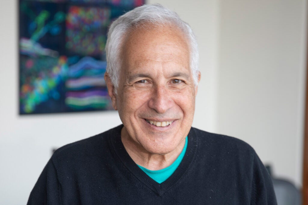
Jeff Lichtman.
Kris Snibbe/Harvard Staff Photographer
The latest map contains never-before-seen details of brain structure, including a rare but powerful set of axons connected by up to 50 synapses. The team also noted oddities in the tissue, such as a small number of axons that formed extensive whorls. Because the sample was taken from a patient with epilepsy, the researchers don’t know whether such formations are pathological or simply rare.
Lichtman’s field is connectomics, which seeks to create comprehensive catalogs of brain structure, down to individual cells. Such completed maps would unlock insights into brain function and disease, about which scientists still know very little.
Google’s state-of-the-art AI algorithms allow for reconstruction and mapping of brain tissue in three dimensions. The team has also developed a suite of publicly available tools researchers can use to examine and annotate the connectome.
“Given the enormous investment put into this project, it was important to present the results in a way that anybody else can now go and benefit from them,” said Google collaborator Viren Jain.
Next the team will tackle the mouse hippocampal formation, which is important to neuroscience for its role in memory and neurological disease.