


Health
Microdevices turn brain tumors into tiny labs
Designed to conduct dozens of experiments at once to study new treatments on deadly brain cancers
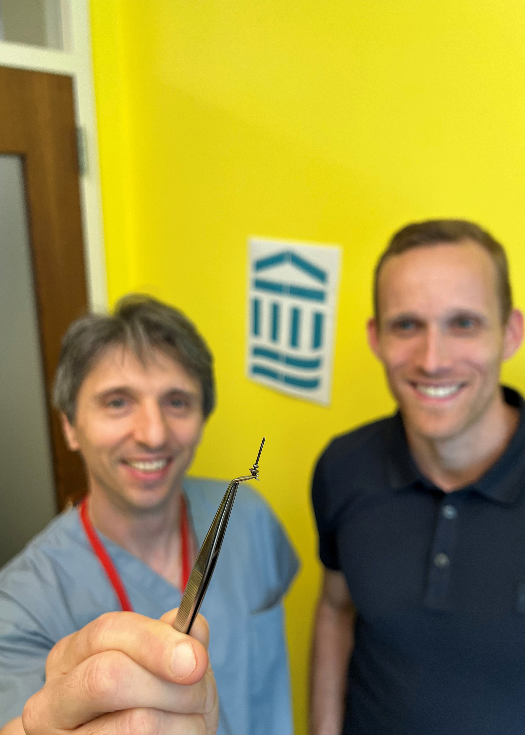
Pier Paolo Peruzzi (left) and Oliver Jonas with the microdevice they designed.
Victoria Roberts/Mass General Brigham Communications
Researchers from Brigham and Women’s Hospital have designed a device that can help test treatments in patients with gliomas, a type of tumor that originates in the brain or spinal cord.
The shape and size of a grain of rice, the new device is designed to be used during standard of care surgery and can conduct dozens of experiments at once offering unprecedented insight into the effects of drugs on glioma tumors.
Results from the pilot clinical trial for the device are published in Science Translational Medicine.
“In order to make the greatest impact on how we treat these tumors, we need to be able to understand, early on, which drug works best for any given patient,” said co-principal investigator and co-corresponding author Pier Paolo Peruzzi, an assistant professor in the Department of Neurosurgery at BWH and Harvard Medical School. “The problem is that the tools that are currently available to answer this question are just not good enough. So we came up with the idea of making each patient their own lab, by using a device which can directly interrogate the living tumor and give us the information that we need,” he explained.
In the time the device is implanted — about 2-3 hours — it administers tiny doses of up to 20 drugs into extremely small areas of the patient’s brain tumor. The device is removed during the surgery and the surrounding tissue is returned to the lab for analysis.
“This is not in the lab, and not in a petri dish. It’s actually in real patients in real time, which gives us a whole new perspective on how these tumors respond to treatment.”
Pier Paolo Peruzzi
Because the device works while the tumor is still in the body, conducting experiments this way gives unparalleled ability to assess the effects of drugs on the tumor microenvironment, the cells immediately surrounding cancer cells that can make up almost half the mass of a tumor.
“This is not in the lab, and not in a petri dish,” said Peruzzi. “It’s actually in real patients in real time, which gives us a whole new perspective on how these tumors respond to treatment.”
About 20,000 people in the U.S. each year are diagnosed with gliomas, a type of tumor that affects the brain and spinal cord. Gliomas are also among the deadliest brain cancers and are notoriously difficult to treat.
One challenge in developing targeted therapies for glioma is that it can be difficult to test many different combinations of drugs in tumor cells, because it’s only possible to treat patients with one approach at a time. This has been a significant barrier for hard-to-treat cancers like gliomas, for which combination therapies are a promising avenue.
Peruzzi worked closely with co-principal investigator Oliver Jonas, an associate professor in the Department of Radiology at BWH, to develop a device that can work around some of the barriers to precision medicine in gliomas. These microdevices are implanted in a patient’s tumor during surgery and removed before the surgery is complete.
“It’s important that we are able to do this in a way that best captures the features of each patient’s tumor and, at the same time, is the least disruptive of the standard of care,” said Peruzzi. “This makes our approach easy to integrate into patients’ treatment and allows its use in real life.”
In the current study, the researchers tested their device on six patients undergoing brain surgery to remove a glioma tumor. None of the patients experienced any adverse effects from the device, and the researchers were able to collect valuable biological data from the devices, such as how the response changes based on drug concentrations, or what molecular changes each drug produces in the cells.
While the study demonstrated that the device was safe and could be easily incorporated into surgical practice, the researchers are still working on determining the exact ways the data it gathers should be used to optimize glioma therapy. The researchers are currently conducting a two-stage version of their procedure in which patients receive the device through minimally invasive surgery 72 hours before their main surgery.
“We’re optimistic that this is a new generation approach for personalized medicine,” said Peruzzi. “The ability to bring the lab right to the patient unlocks so much potential in terms of the type of information we can gather, which is new and exciting territory for a disease that has very few options at present.”
Disclosures: Pier Paolo Peruzzi is co-founder and on Board of Directors of Ternalys Therapeutics. He is a named inventor on microRNA-related patent PCT/US2019/029988. Jonas is a co-inventor of the IMD technology (U.S. patent no. US10390702B2) and is a consultant with compensation (non-equity) to Kibur Medical. Co-author Antonio Chiocca is a named inventor on patents related to oncolytic HSV1 (U.S. patent nos. US10,806,761B2 US10,232,002B1, and US2017/0015757A1) and noncoding RNAs (PCT/US2019/029988).
This study was supported by grants from the National Institutes of Health (R37CA224144, P41EB028741) and the National Institute of Neurological Disorders and Stroke (1R01NS116144).