


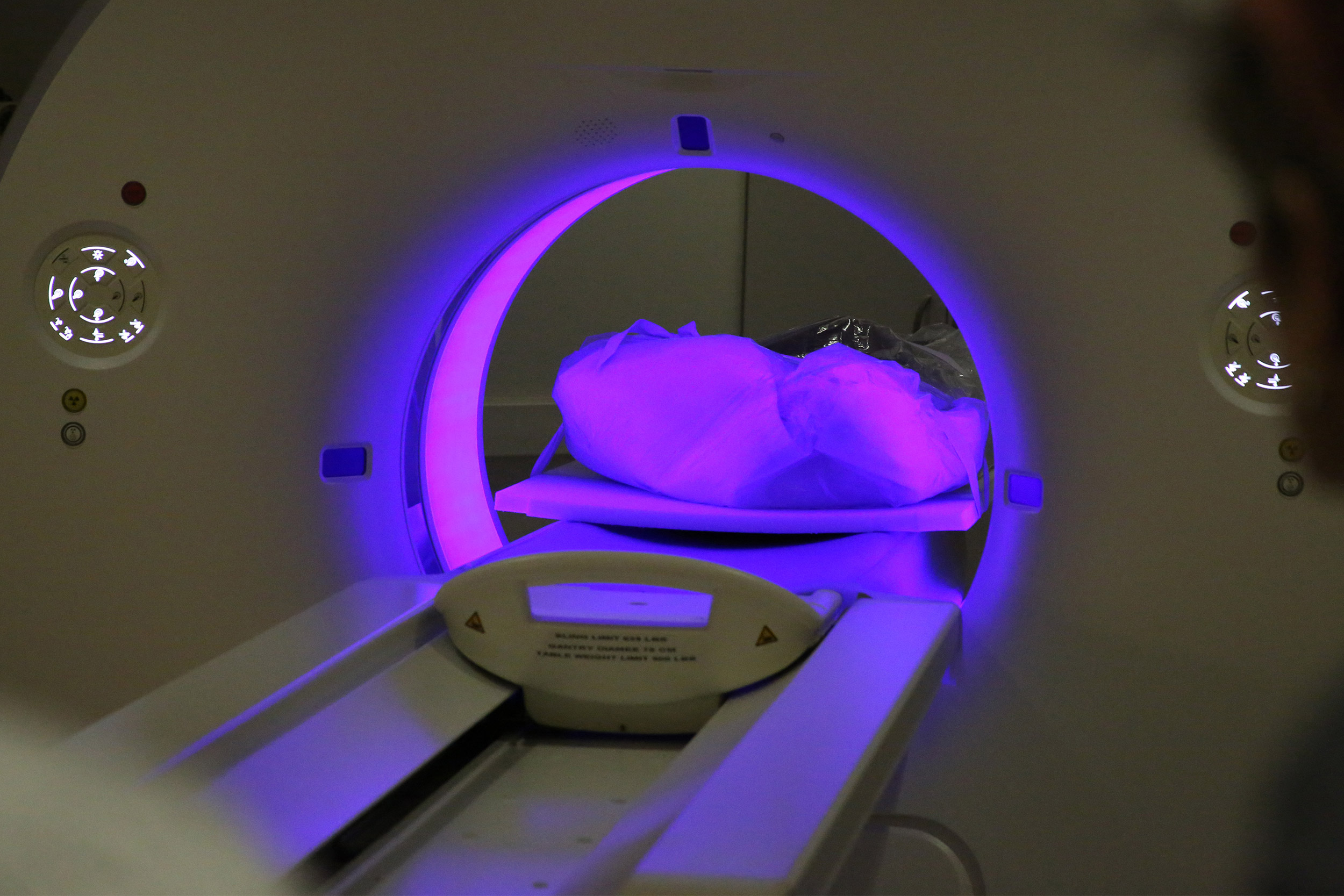
High-resolution scans of the mummified remains revealed the telltale hardened calcium deposits in various blood vessels in the chest.
Credit: Brigham and Women’s Hospital
Science & Tech
Clues of heart disease found in 16th-century mummies
Despite ‘heart-healthy’ diet, researchers discover evidence of plaque
What secrets lie in the hearts of our ancestors? Signs of cardiovascular disease, for one, as a team of cardiovascular-imaging experts from Brigham and Women’s Hospital (BWH) recently helped discover.
Through a collaboration with an international team of researchers and anthropologists, BWH faculty and staff performed CT scans on five mummies from 16th-century Greenland in the Shapiro Cardiovascular Center early last year. The team was looking for evidence of plaque in the arteries — also known as atherosclerosis — to see if the leading cause of death in the U.S. today was also prevalent centuries ago.
Sure enough, high-resolution scans of the mummified remains — belonging to four young adults and one child from an Inuit community — revealed the telltale hardened calcium deposits in various blood vessels in the chest.
“It’s always fascinating to look at humans who lived hundreds of years ago and see if learning about the past could teach us more about the present and future,” said Ron Blankstein, associate director of the Brigham’s Cardiovascular Imaging Program, director of cardiac computed tomography, and a preventive cardiology specialist.
Blankstein was among the experts who scanned the mummies and interpreted the images in 2018, an event featured on National Geographic’s “Explorer” series. The effort was part of a broader project, led by a group of external researchers, to scan mummies from hunter-gatherer and preindustrial civilizations around the world to search for signs of heart disease.
Searching the past
From Egypt to Mongolia and now Greenland, mummies throughout the ages have shown evidence of atherosclerosis. The Greenland mummies were of particular interest due to their diet, which would have primarily consisted of fish and sea mammals.
While increased fish consumption is commonly touted as heart-healthy — which may make the findings of atherosclerosis seem surprising — Blankstein emphasized that scientists still have much to learn about its relationship to cardiovascular health. For example, although it is known that consuming fish rich in omega-3 fats has benefits, some types of fish can also be high in cholesterol and, in the current era, contain toxins like mercury or polychlorinated biphenyls (PCBs) that may pose risk, he said.
Lifestyle factors, such as exposure to cooking smoke in their dwellings, may have also contributed to the mummified individuals developing cardiovascular disease during their lifetimes, Blankstein said. Given that and the small sample sizes of these mummy scans, he noted that the team’s findings shouldn’t be taken too much to heart.
“The question of whether fish is good or bad for you is still open-ended, and it would be unrealistic to think that we could provide a definitive answer by scanning a small number of mummies for plaque,” Blankstein said. “Our team found it fascinating that there was evidence of atherosclerosis despite the mummies’ estimated young ages, but this also doesn’t mean cardiovascular disease is inevitable. In fact, the majority of cardiovascular disease events that we see in patients is preventable with appropriate diet, weight control, and lifestyle changes, such as regular exercise; at times, medication can also be used to treat various risk factors.”
A different kind of patient
Scanning the mummies wasn’t too different from work the cardiovascular imaging team normally does. In fact, they were a little easier to scan than living patients; normally, the CT scanner must account for the movement of a beating heart.
Interpreting the images required a different perspective, however, Blankstein explained.
“This is not the same as scanning a [living] human. All of the organs are decomposed — in fact, you don’t see much of the heart at all,” he said. “The major plaque we saw was not necessarily in the arteries of the heart but in some other blood vessels in the chest, such as the aorta or some arteries of the neck.”
In addition to satisfying the team’s intellectual curiosity, Blankstein said he hopes the findings will inspire people to learn more about atherosclerosis and how to reduce their risk.
Other faculty and staff who helped conduct the scans included Kristen Burke, CT technologist; Marcelo Di Carli, director of the Cardiovascular Imaging Program and chief of the Division of Nuclear Medicine; Abe Haboub, cardiac CT manager; and Michael Steigner, director of vascular imaging.
To read the full story, visit the Brigham Bulletin website.


