
Science & Tech
Chilling discovery
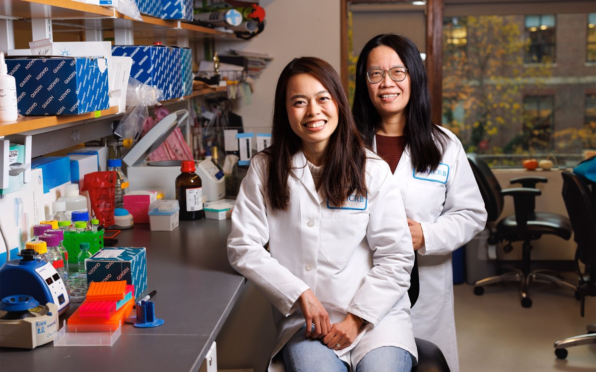
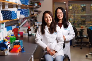
Suk Hyun Sung (left) and Ismail El Baggari.
Photos by Niles Singer/Harvard Staff Photographer
Physicists go to extremes to capture quantum materials
Researchers at the Rowland Institute at Harvard have pioneered a new way to achieve the coolest possible temperatures to image materials at sub-atomic scale. In combining the technical know-how of Rowland staff scientists with collaborators at the University of Michigan, the findings make possible a new era of ultra-cold microscopy.
Cryogenic transmission electron microscopy — TEM — has long played a vital role in many branches of science, from biology to physics, because the very low temperatures allow close examination of samples of everything from inorganic crystals to complex biomolecules at the atomic scale. Typically, the cryogen, or cooling agent, is liquid nitrogen, which boils at 321 below zero Fahrenheit (or 77 Kelvin) — impressive, but not cold enough to see those strange quantum wriggles.
That’s why scientists have strived over the last decade to go colder by using liquid helium, which boils at 421 below zero Fahrenheit, or 4 Kelvin, and is very close to “absolute zero.” But this comes with serious technical problems that affect the mechanical stability of the microscope.
Harvard researchers envisioned a new way to use liquid helium for a more stable approach to this high-level microscopy, a breakthrough explained in a paper published last month in PNAS. Led by principal investigator and Rowland fellow Ismail El Baggari, the team built novel partnerships to create a usable new technology, both within the institute and outside, with University of Michigan Professor Robert Hovden. The research is among the first high-impact papers (another was robot flies) since Rowland moved to Harvard’s science campus a year ago.
The original concept of cryogenic cooling during microscopy to preserve the structure of biological molecules — which led to the 2017 Nobel Prize for chemistry — was to use it to study cells, proteins, and other soft matter. However, El Baggari and Suk Hyun Sung, a postdoc on the team, saw its potential in quantum physics.
“We’re physicists and we’re interested in imaging weird materials called quantum materials that have different properties at low temperatures,” said El Baggari, noting that electrons exhibit quantum behavior, only at extremely low temperatures. “And so we need to cool down the materials to access those new properties.” Liquid helium cooling was seen as a means to do this.
To utilize liquid helium, however, the researchers had to have an electron microscope that would allow such cooling. “In the past, we’ve used electron microscopy to image the atomic structure of these materials at high temperatures, but it turns out that it’s very difficult to get high-resolution images with liquid helium cooling,” El Baggari said.
“Liquid helium evaporates so quickly and so easily any external heat vaporizes it,” he added. “That then introduces vibrations as bubbles form, as the flow is disturbed by a mixture of gas and liquid.”
These vibrations blurred the images in the way a bubbling glass saucepan of boiling water obscures the view through the pot, though that’s visible when the water is cold. The fast evaporation also meant that the cold temperatures could be maintained for only about 20 minutes, far too short for meaningful measurements.
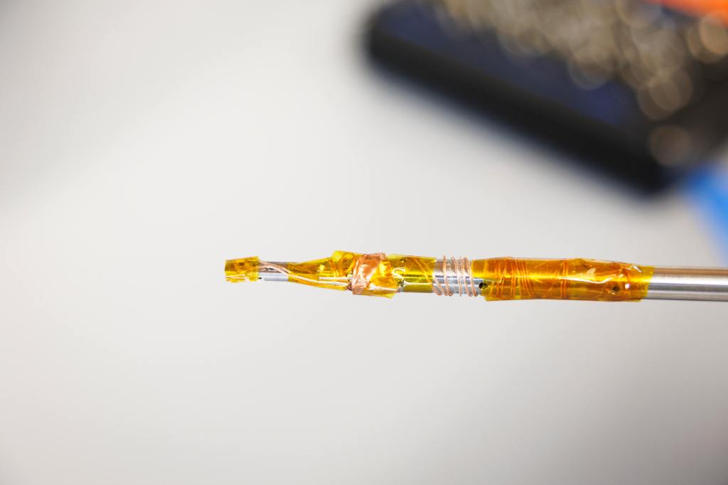
“We built a geophone for the microscope that is so sensitive, it can detect vibrations within fractions of atomic distance.”
Winfield Hill, director of electronic engineering
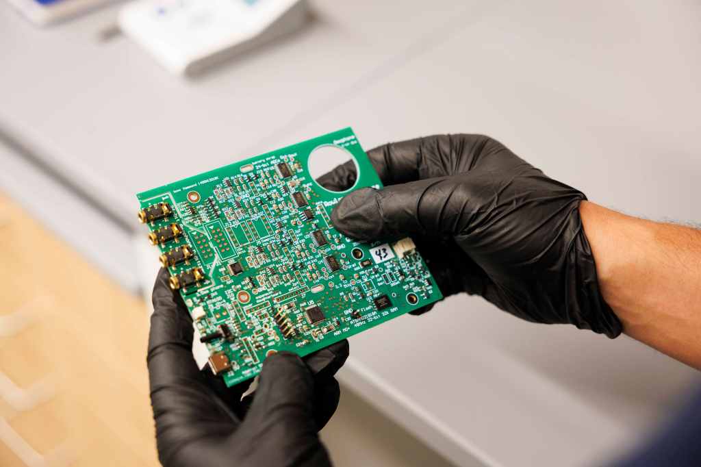
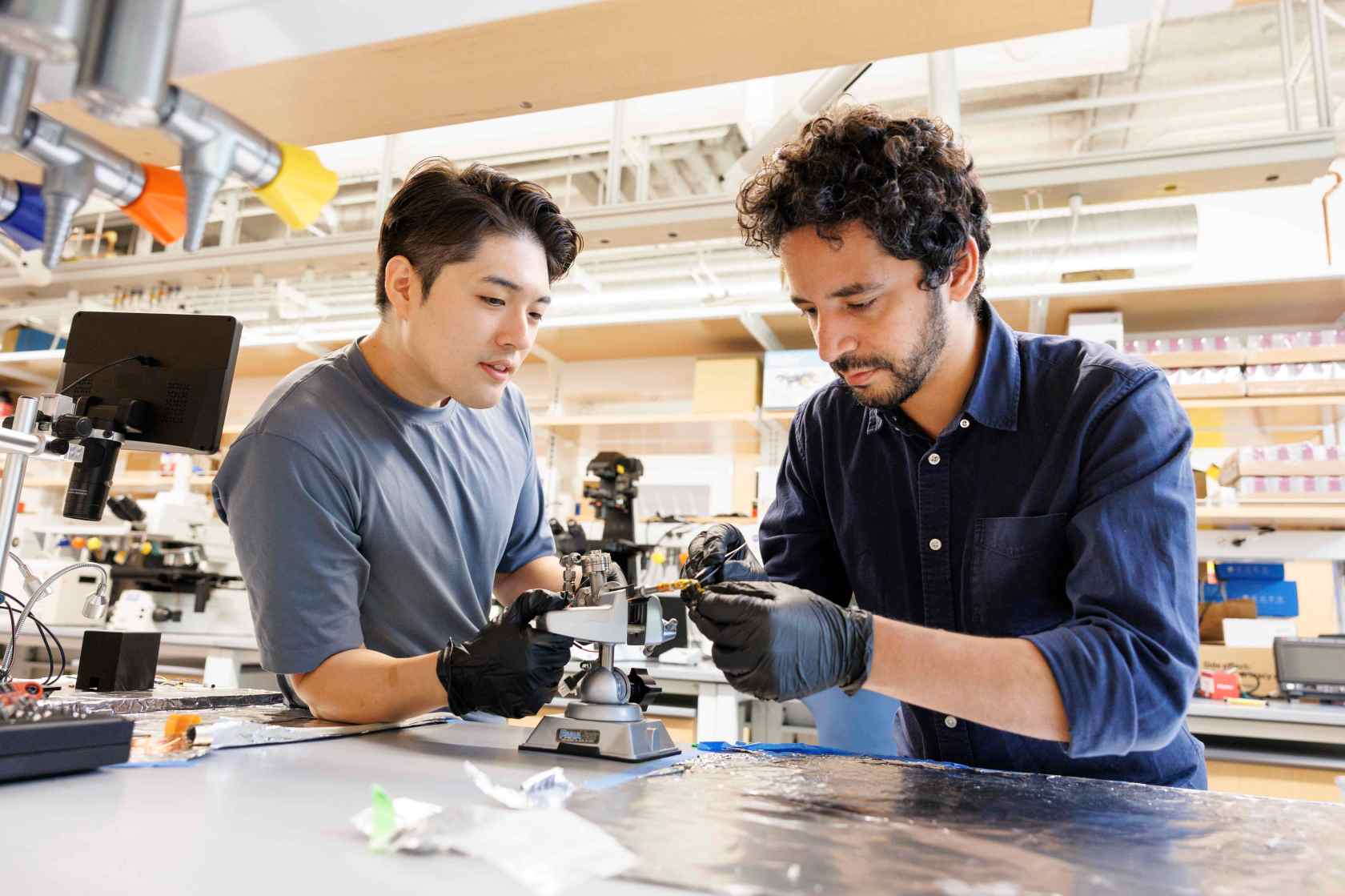
To solve this problem, the researchers needed to create a method that would allow the use of liquid helium, but in a way that didn’t allow vibrations. The researchers collaborated with Rowland manager Erik Madsen; director of electronic engineering Winfield Hill; and Alan Stern, a staff computational scientist. Together, they built a system that could maintain cold temperatures for hours on end. They also tracked and isolated the vibrations of the cooling system from reaching the specimen.
“We had the general ideas, but we didn’t know at the start the details of cryogenic design, machining to high tolerances, and precision electronics,” said El Baggari. “We had to develop new tools and processes to begin tackling our problems.”
“Ismail had the idea to use geophones to detect minuscule vibrations in the ultra-cold TEM set-up, up to identify places to dampen the ‘noise,’ particularly on the liquid helium input. So we built a geophone for the microscope that is so sensitive, it can detect vibrations within fractions of atomic distance — exactly the scale needed for this microscope to be able to image clearly at sub-atomic resolution,” said Hill.
The stakes were high because of the complexity of the machines. “These microscopes are multimillion-dollar machines, and it was quite nerve-wracking to think about inserting our own new instrument in what has never been tested,” said Sung.
Into this huge and delicate machine, he continued, the builders had to create site entry holders that “would fit in like a glass slide fits into an ordinary microscope — without breaking anything.” The sample had to land in the right position of within 25 microns, or a third of the width of human hair. Any deviations meant the sample could not be imaged, vibrations were excessive, or vacuum levels were poor.
“For the first months of building things, we were just measuring commercially available site entry holders, making sure that all the dimensions were correct,” continued Sung. “We didn’t know what the critical dimensions were because all the holders were slightly different, and we didn’t know what actually mattered.” After three years of testing the sample holder and improving vibration isolation, the team was able to capture the first high-resolution images in quantum materials while operating for many hours at ultracool temperatures.
“Suddenly we were studying materials that we historically had only been able to examine at room temperature,” said El Baggari. “We were never able to image them while they were actually exhibiting the emergent properties that we and our collaborators are interested in. So now we can actually use the electron microscope as a primary tool to directly tackle these quantum electronic phases in materials.”
Continued improvements of the new tool could open new fields of inquiry, such as simultaneously applying a voltage to working devices or manipulating materials by stretching or pressing on them. “The microscopy experiments that we’ve been routinely doing at room temperatures, we can now do it at low temperatures where things get even more exciting,” he said.
The research described in this article was partially funded by the National Science Foundation and the U.S. Department of Energy.