
Campus & Community
Dusting off a microscopic portion of Harvard’s Glass Flowers collection
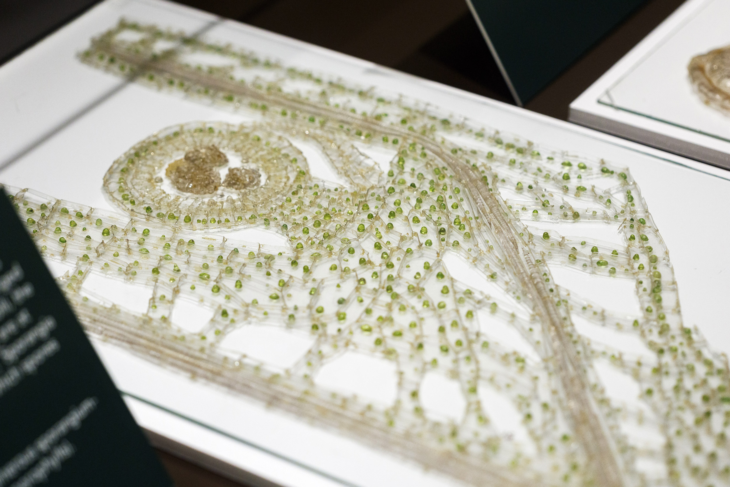
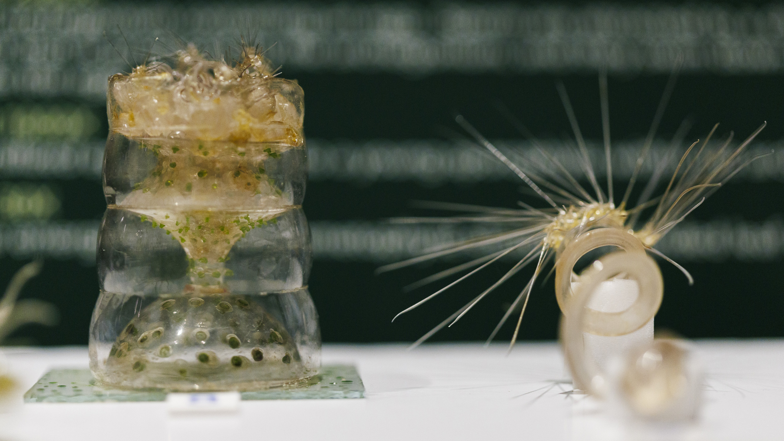
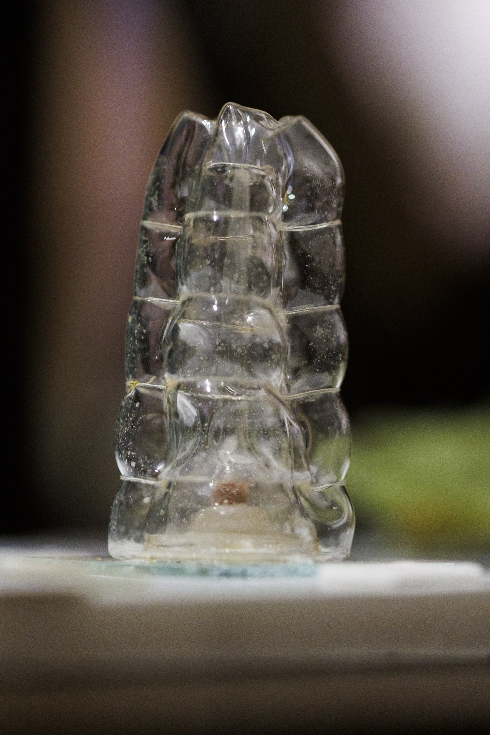
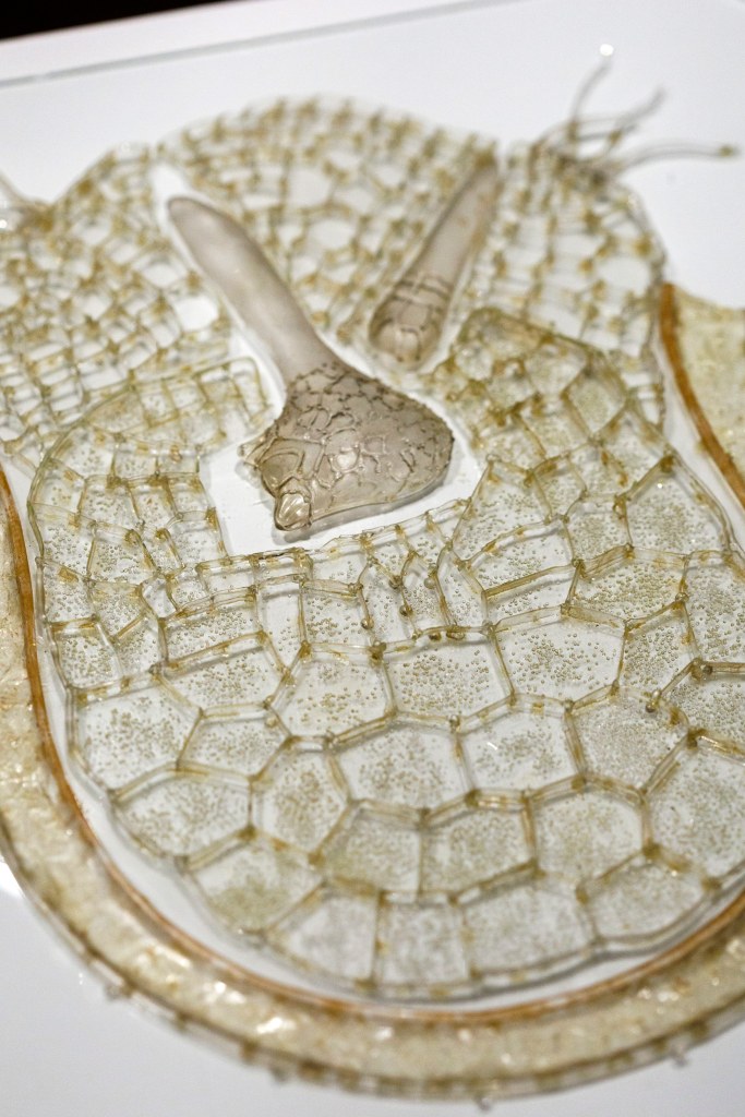
“The Blaschkas at the Microscope: Lessons in Botany” in the Glass Flowers gallery features models crafted by Leopold and Rudolf Blaschka that illustrate life cycles of spore-forming plants and fungi. A section through the stem, spore-bearing container, and leaf of the spike-moss, Selaginella sp., magnified 500X.
Photos by Stephanie Mitchell/Harvard Staff Photographer
New release shows minute details of lives of spore-forming plants and fungi
Models created more than 100 years ago may leave some viewers wondering which is more miraculous, the original or the replica?
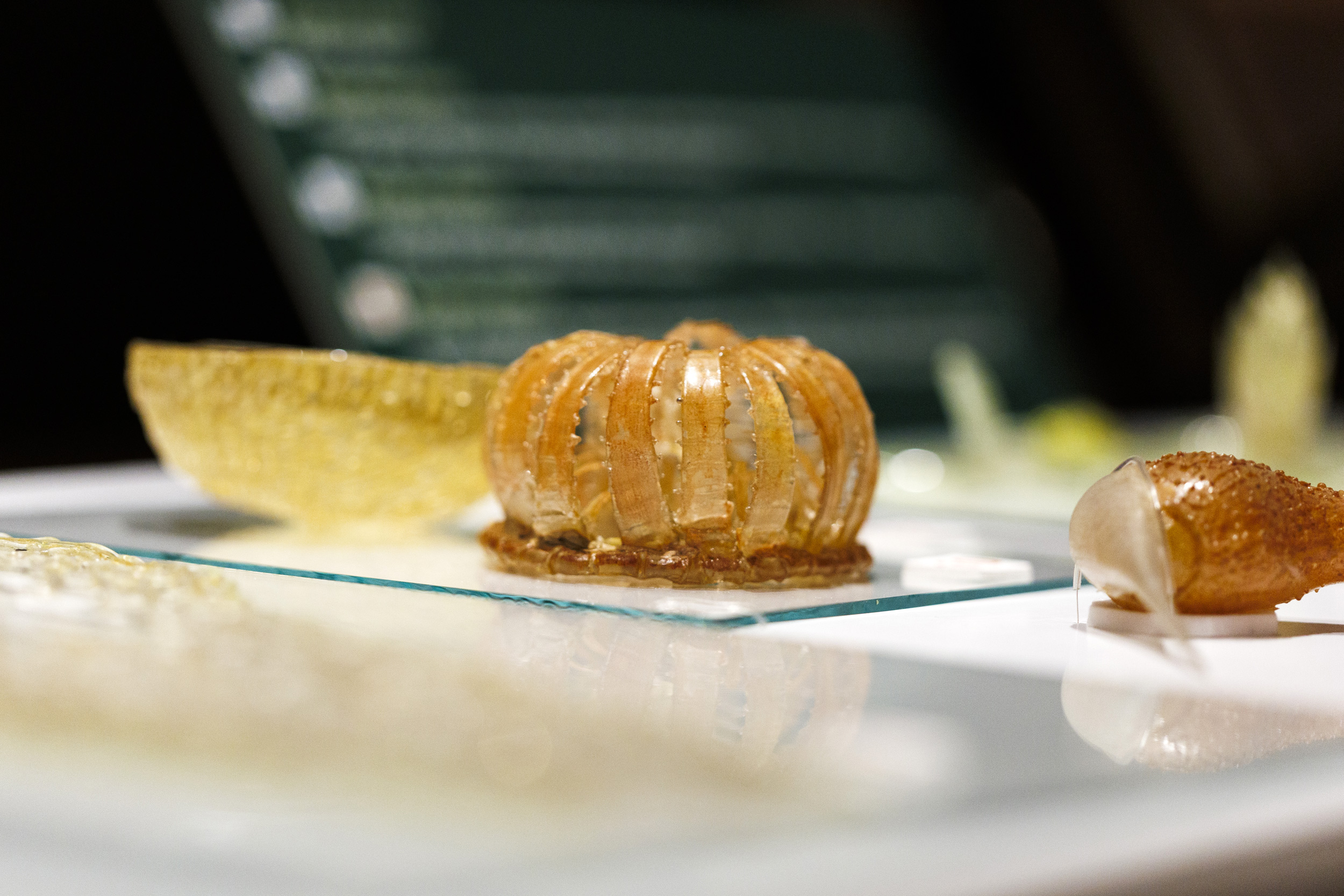
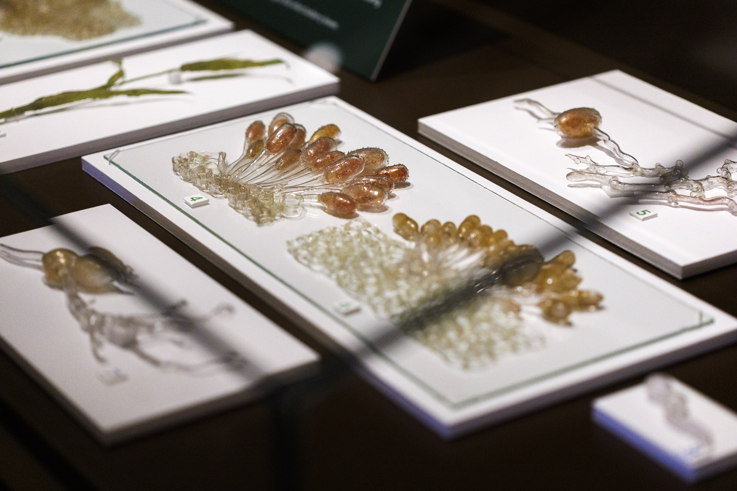
Since April, “The Blaschkas at the Microscope: Lessons in Botany” at the Harvard Museum of Natural History has showcased a series of models produced between 1889 and 1893 by father-and-son of Czech glass artists Leopold (1822-1895) and Rudolf Blaschka (1857-1939). Between 1886 and 1936, the Blaschkas produced, exclusively for Harvard, 4,300 exquisite glass models representing 780 plant species, tropical and temperate, in various stages of health and disease.
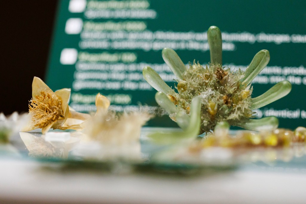
The current exhibit, exploring microscopic details of the life cycles of spore-forming plants and fungi, puts on display models produced between 1889 and 1893 that have not been seen in nearly a quarter century. It also explains how mosses survive prolonged periods of dehydration, and how ferns have survived to become one of the oldest plant groups on Earth, and explores pathogens that threaten the survival of all organisms.
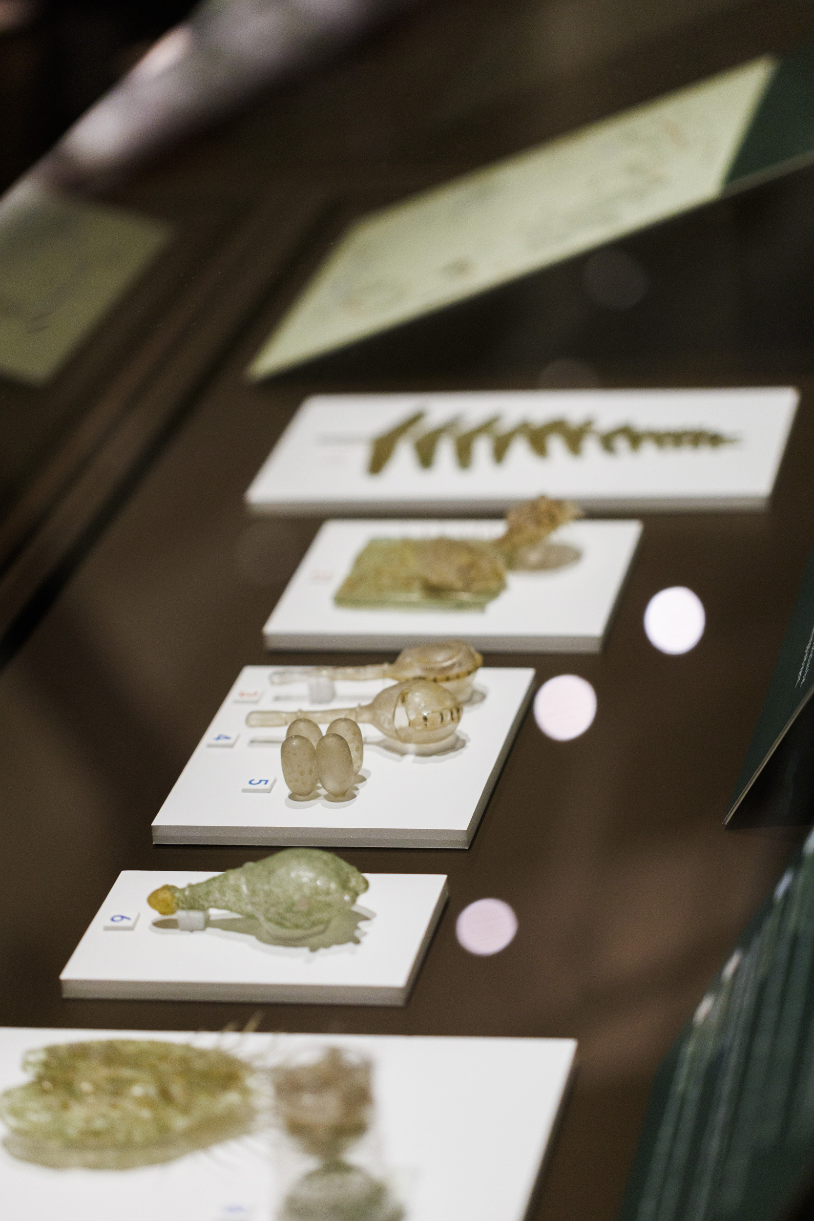
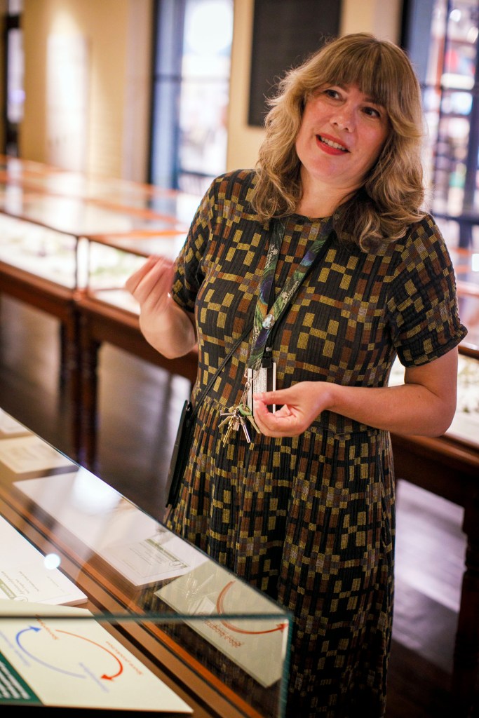
“This special exhibition illustrates and explains these complex life cycles to visitors through the glass models, carefully written text, and accompanying diagrams,” said Jennifer Brown, collection manager of the Ware Collection of Blaschka Glass Models of Plants, Harvard University Herbaria. “The models also show another aspect of the Blaschkas’ artistry. The extreme magnifications rendered in glass-on-glass plates are incredible.”
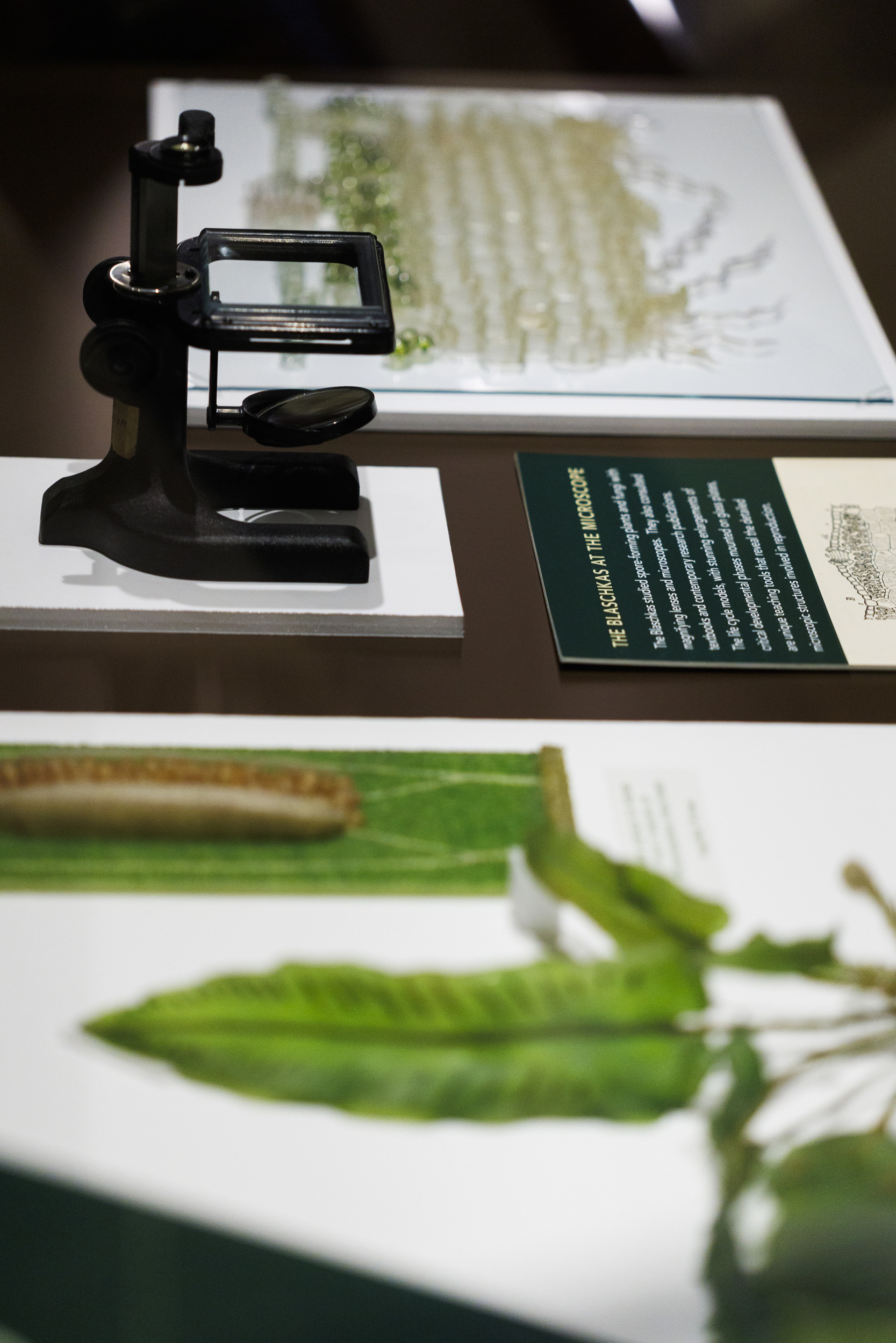
The Blaschkas used their own observations, dissections, botanical textbooks, magnifying lenses, and microscopes to create accurate models of the reproductive cycles of ferns, mosses, liverworts, and pathogenic fungi. Their enlargements of microscopic structures are critical to understanding plant and fungal reproduction.
The exhibition was curated by Michaela Schmull, director of collections, Harvard University Herbaria, and Donald H. Pfister, Asa Gray Research Professor of Systematic Botany and curator of the Farlow Library and Herbarium, Harvard University Herbaria, Emeritus. Scott Fulton, conservator of the Ware Collection of Blaschka Glass Models of Plants, was responsible for making sure the models look their best after being in storage for almost 25 years.
The exhibit runs through February 2026.