How the gut got its villi
“You are not just a ball of cells,” says Clifford Tabin, George Jacob and Jacqueline Hazel Leder Professor of Genetics at Harvard Medical School (HMS).
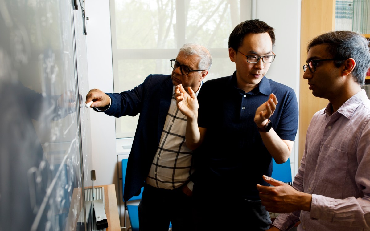
The way cells organize within the human body allows us all to function the way we do, but a couple of Harvard professors are concerned as much with that developmental process as with the end result. Tabin shares a common perspective with L. Mahadevan, the Lola England de Valpine Professor of Applied Mathematics at the Harvard School of Engineering and Applied Sciences (SEAS), professor of organismic and evolutionary biology, and professor of physics.
“When I teach medical students, they’re more interested in the rare people who are born with birth defects,” says Tabin. “They want to understand embryology so they understand how things go awry, but I’m more interested in the fact that for everyone sitting in my classroom—all 200 of those medical students and dental students—it went right! And every one of them has a heart on the left side and every one of them has two kidneys, and how the heck do you do that?”
By taking steps back through embryos’ development, researchers in Mahadevan’s and Tabin’s laboratories investigated how the guts of several different animals end up as they do. Their findings, published in a recent issue of Science, reveal that the principles guiding the growth of intestinal structures called villi are surprisingly similar across chickens, frogs, mice, and snakes.
