


iStock by Getty Images
Science & Tech
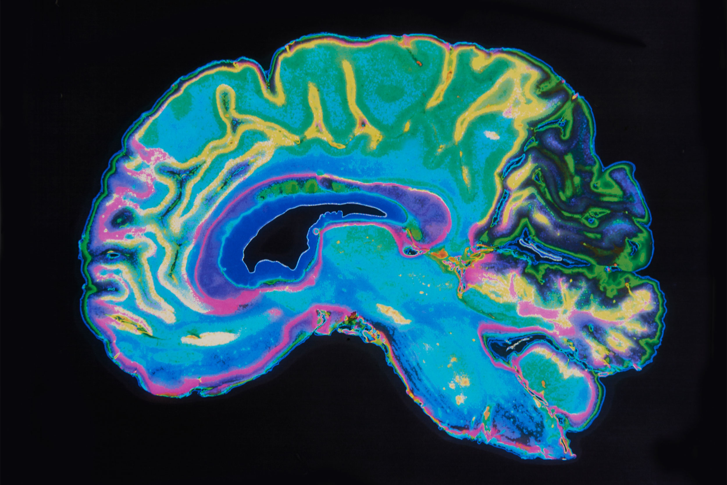
Cellular atlas guides new understanding of brain
Innovative technology gives voice to pathologic changes in neurodegenerative illnesses like Alzheimer’s
By combining noninvasive imaging techniques, investigators have created a comprehensive cellular atlas of a region of the human brain known as Broca’s area — an area critical for producing language.
The new technology will provide insights into the presence and spread of pathologic changes that occur in neurodegenerative illnesses — such as epilepsy, autism, and Alzheimer’s disease — as well as psychiatric illnesses.
Until now, scientific advances have not produced undistorted 3D images of cellular architecture that are needed to build accurate and detailed models. In new research published in Science Advances, a team led by investigators at Harvard-affiliated Massachusetts General Hospital, has overcome this challenge with detailed resolution to study brain function and health.
Using sophisticated imaging techniques — including magnetic resonance imaging, optical coherence tomography, and light-sheet fluorescence microscopy — the researchers were able to prevail over the limitations associated with any single method to create a high-resolution cell census atlas of a specific region of the human cerebral cortex, or the outer layer of the brain’s surface. The team created such an atlas for a human postmortem specimen and integrated it within a whole-brain reference atlas.
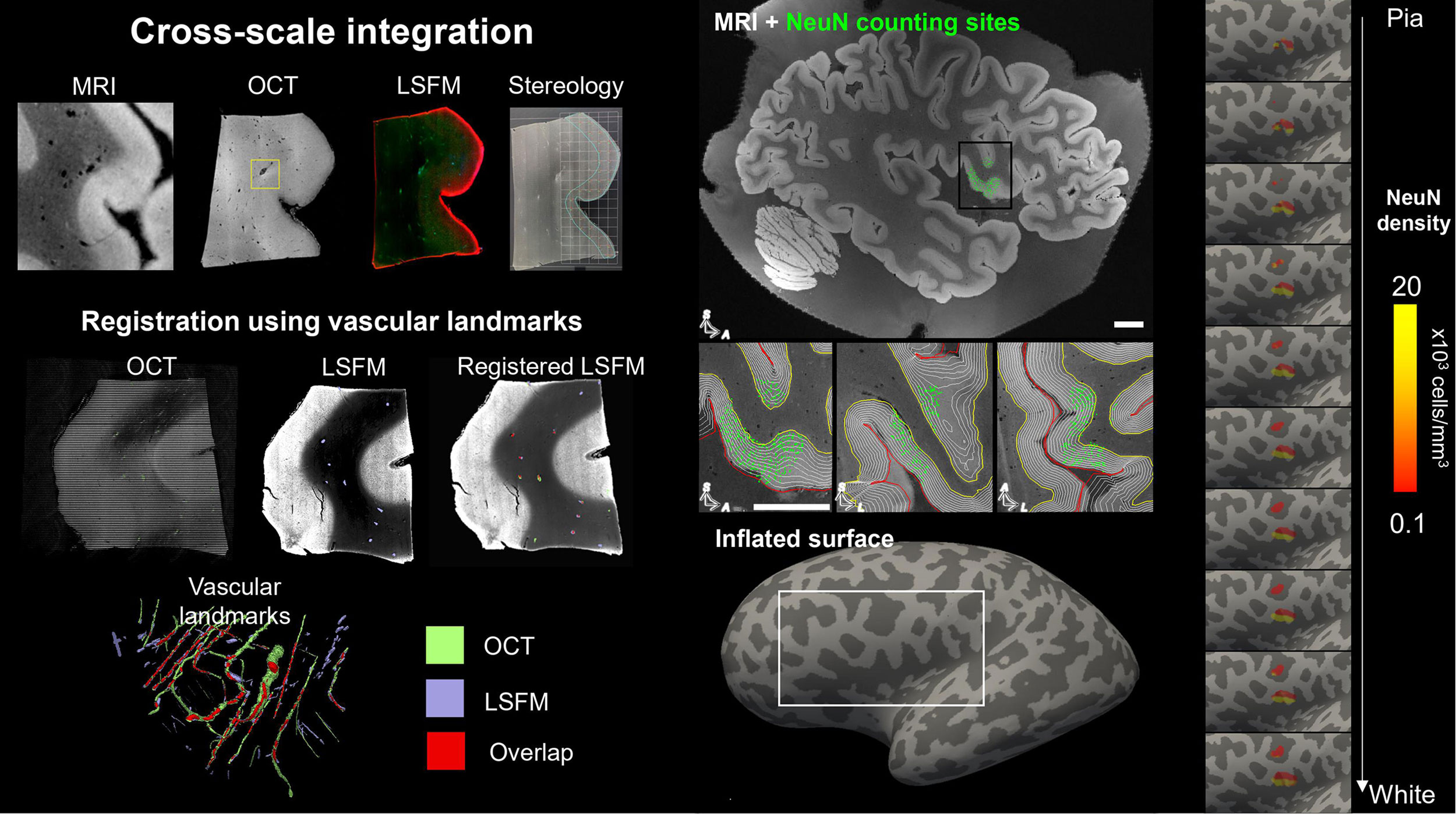
Overview of the new pipeline. Human brain samples are imaged at multiple scales with multiple modalities (MRI, OCT, and light-sheet fluorescent microscopy or LSFM).
MGH
“We built the technology needed to integrate information across many orders of magnitude in spatial scale from images in which pixels are a few microns to those that image the entire brain,” says co–senior author Bruce Fischl, director of the Computational Core at the Athinoula A. Martinos Center for Biomedical Imaging at MGH and a professor in radiology at Harvard Medical School.
Ultimately, the methods in this study could be used to reconstruct undistorted 3D cellular models of particular brain areas as well as of the whole human brain, enabling investigators to assess variability between individuals and within a single individual over time.
“These advances will help us understand the mesoscopic structure of the human brain that we know little about. Structures that are too large and geometrically complicated to be analyzed by looking at 2D slices on the stage of a standard microscope, but too small to see routinely in living human brains, says Fischl.
“Currently we don’t have rigorous normative standards for brain structure at this spatial scale, making it difficult to quantify the effects of disorders that may impact it such as epilepsy, autism, and Alzheimer’s disease.”
Additional co-authors include Irene Costantini, Leah Morgan, Jiarui Yang, Yael Balbastre, Divya Varadarajan, Luca Pesce, Marina Scardigli, Giacomo Mazzamuto, Vladislav Gavryusev, Filippo Maria Castelli1, Matteo Roffilli, Ludovico Silvestri, Jessie Laffey, Sophia Raia, Merina Varghese, Bridget Wicinski, Shuaibin Chang, Anderson Chen I-Chun, Hui Wang, Devani Cordero, Matthew Vera, Jackson Nolan, Kimberly Nestor, Jocelyn Mora, Juan Eugenio Iglesias, Erendira Garcia Pallares, Kathryn Evancic, Jean Augustinack, Morgan Fogarty, Adrian V. Dalca, Matthew Frosch, Caroline Magnain, Robert Frost, Andre van der Kouwe, Shih-Chi Chen, David A. Boas, Francesco Saverio Pavone, and Patrick R. Hof.
Support for this research was provided in part by the BRAIN Initiative Cell Census Network, the National Institute for Biomedical Imaging and Bioengineering, the National Institute on Aging, the National Institute of Mental Health, the National Institute for Neurological Disorders and Stroke, Eunice Kennedy Shriver National Institute of Child Health and Human Development, Chan-Zuckerberg Initiative DAF an advised fund of Silicon Valley Community Foundation, Shared Instrumentation, NIH Blueprint for Neuroscience Research, European Union’s Horizon 2020 research and innovation Framework Programme, European Union’s Horizon 2020 Framework Programme for Research and Innovation, Marie Skłodowska-Curie, Italian Ministry for Education in the framework of Euro-Bioimaging Italian Node, European Research Council, Alzheimer’s Research UK, The National Institutes of Health, and “Fondazione CR Firenze” (private foundation) Human Brain Optical Mapping.


