
Science & Tech
Think of them as utility players
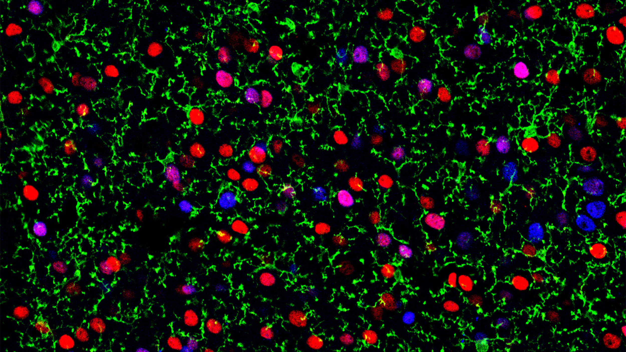
Microglia (in green) in a sea of different excitatory neuron types (colored red or blue). A new study shows how microglia are prompted to change their state to adapt to different areas of the brain.
Credit: Arlotta lab
New study shows how tiny immune cells are prompted to change their state to help neurons with brain function
To accomplish feats of athletic or academic excellence and even everyday actions like walking and talking, the brain must acquire and seamlessly process a ton of information. That requires a whole orchestra of cells to “listen” and move, interact, and coordinate with one another. One of the most enduring, fundamental questions in neuroscience involves precisely how this happens.
Scientists know that this cellular symphony includes not only neurons, but cells that normally play a role in defending the body against pathogens. One group is tiny immune cells called microglia, which researchers are increasingly learning play oversized roles in brain function, health, and disease. The cells are also gaining increased attention for their roles in assembling and maintaining neural circuits and their ability to change their molecular identity to match their environment. A key to solving one of neuroscience’s great mysteries involves finding out how they make this change.
In a new report in Nature, researchers from the lab of Paola Arlotta, Golub Family Professor of Stem Cell and Regenerative Biology and the Stanley Center for Psychiatric Research at the Broad Institute, move a step closer to answering this question. The paper, published Wednesday, shows that microglia cells “listen in” to neighboring neurons and change their molecular state to match them.
“When they were first discovered, microglia were assumed to be simply scavengers, cleaning up cell debris and helping to fight off pathogens,” said Jeffrey Stogsdill, who led the study as a postdoctoral researcher in the Arlotta lab. “Now we know that microglia can interact with neurons in very sophisticated ways that can affect neuron function.”
“We know that microglia can affect the function of the neural circuit, and now we know that neurons can recruit specific types of microglia to their neighborhood.”
Paola Arlotta
This discovery could one day open the door for lines of research that can target communications between microglia and their neuron partners with pinpoint accuracy, offering insight into disorders such as autism and schizophrenia, which arise when these communications between cells go awry.
“You would no longer have to treat, for instance, microglia as one blanket cell type when trying to affect the brain,” Stogsdill continues. “We can target very specific states, or we can target very specific subtypes of neurons with the ability to change specific states of microglia. It allows us to have high-level granularity.”
The study provides unique insight into how different cell types work together in harmony.
“What we’re discovering here are the rules by which different cell types in the brain talk to each other and influence each other to ultimately be able to do more together,” said senior author Arlotta, an institute member at the Broad.
In the paper, the scientists describe how neurons train microglia to work with them when they first meet, early in the life of the brain. The group found that during the formation of the cerebral cortex — a part of the brain responsible for skilled motor function, sensory perception, and cognition — different types of neurons influence the number and molecular state of nearby microglia in their own unique ways.
“These different types of cortical neurons recruit different numbers of microglia,” Stogsdill said. “They then pattern those microglia to tell them exactly what type they need to be.”
The cerebral cortex is organized into layers, with different neuron types residing in each. The researchers used genetic profiling methods to examine the microglia in the different layers and discovered that they varied in number and molecular state depending on the layer in which they were found. The team then changed the composition of neuron types in these layers and found that they could influence the distribution of the different microglial states. The microglia matched the types of neurons in the new locations, confirming that the neurons were influencing them.
The research team then built a molecular atlas that outlined the communication between neurons and microglia. The team analyzed their profiling data to find pairs of interacting proteins expressed by the different microglial states and their neuron partners. Such a molecular atlas could enable future research into the functional roles of these interactions and possible targets for therapeutic intervention. The researchers plan to start by explaining exactly what differences and functional distinctions exist among microglia in the different layers.
“We know that microglia can affect the function of the neural circuit, and now we know that neurons can recruit specific types of microglia to their neighborhood,” Arlotta said. “It’s a fascinating idea that neurons can reshape their environment to help fine-tune their own circuit function.”
This work was supported by grants from the Broad Institute of MIT and Harvard, the U.S. National Institute of Health, the Klarman Cell Observatory, and the Howard Hughes Medical Institute.