


Campus & Community
Beyond Phineas Gage
‘Oddities’ at HMS’s Warren Museum are also valuable research tools
Skeletons of conjoined twins and legs corkscrewed with rickets. Kidney stones the size of golf balls. The skull of a man who survived a crowbar shot through his head.
The Warren Museum at the Harvard Medical School (HMS) knows how to get your attention. But the value of the museum, which recently reemerged after several years in hiding, goes far beyond the gross-out factor. “There’s actual valid medical, valid historical, valid contemporary use for the collection,” says curatorial manager Virginia Hunt, who is on a crusade to make over the museum’s image.
Established at the Medical School in 1847, when John Collins Warren donated his personal teaching collection of anatomical models to the school, the museum is one of the few medical museums founded in the 19th century that still exists. The museum’s collection was housed in Gordon Hall for most of the 20th century until it slipped gradually into storage throughout the 1990s. By 1998, the museum’s 15,000 specimens were entirely invisible, stored in warehouses off-site.
In September 2000, the museum resurfaced as part of the Countway Library Rare Books and Special Collections Department, displaying 300 of its “greatest hits” in a thoughtful and thought-provoking exhibit on the library’s fifth floor. Now Hunt and collections manager Suzanne Fitz are cataloging the collection, updating century-old paper records into a searchable database that they hope will help entice researchers to mine the museum’s rich resources.
Tools to train doctors
Modern medicine was a new field when Professor Warren, best known for his involvement in the first demonstration of ether anesthesia at Massachusetts General Hospital in 1846, donated the anatomical teaching models that formed the backbone of the collection. The models are human cadavers, dried and coated with resin, the tissue dissected, the veins and arteries injected with colored wax. Warren also donated wet tissue specimens, which are in storage.
“He created the collection because he wanted his students to see what normal anatomy looked like,” says Hunt, who notes that social taboos about dead bodies hindered medical study in Warren’s day. Until 1830, it was illegal to dissect bodies for anatomical teaching in Massachusetts.
The museum provides many such glimpses into the history of medicine and its study. Hunt calls the instrument collection the “oh my God, this is how they used to practice medicine stuff.” There are amputation kits that look like they belong in a carpenter’s tool chest and showy lancet cases and surgery tools of gold and ivory – beautiful, but hardly hygienic. Some items, like early stethoscopes and obstetric forceps, are remarkable in their similarity to today’s tools.
Phineas Gage’s crowbar skull
While Warren’s models, and other models of wax and papier mâché, focus on normal anatomy (the workaday stuff for Victorian-era medical students) later collectors brought abnormal specimens – and gawkers – into the Warren Museum. Headlining at the hall of human horrors is the “crowbar skull” of New Hampshire construction foreman Phineas Gage.
Gage, by far the Warren’s most famous exhibit, won this dubious notoriety working on the Rutland & Burlington Railroad in Cavendish, Vt., when, in September of 1848, an accidental explosion fired a 13-pound tamping iron through his head. The three-foot rod, which accompanies Gage’s skull in the exhibit, landed several feet away from Gage.
Remarkably, Gage survived the horrendous accident, which tore his scalp and fractured his skull. But when he returned home after 10 weeks, it was clear that the once well-liked Gage had undergone a dramatic change in personality. He had become obstinate and impatient; friends described him as “no longer Gage.”
Gage unwittingly ushered in new thinking about personality and the brain. Previous studies of personality had been based on phrenology, which analyzed personality traits based on the size and shape of one’s skull. Gage’s accident turned the focus toward the physiology of the brain.
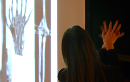
New research from old bones
At a recent lecture at the Warren, HMS tutor Peter Ratiu presented new research on Phineas Gage’s 150-year-old injury. As the museum regains its public presence, Hunt hopes that more researchers like Ratiu will take advantage of the collection. “It’s a wonderful key to the practice of medicine and thoughts about health at the time,” she says. From anthropologists and historians to forensic scientists, radiologists, and geneticists (who can take DNA from the wet tissue and bone specimens) the researchers who might benefit from the Warren’s collection are many.
Collections manager Fitz, who has a background in forensics herself, points out some disturbingly timely applications for the collection: the Warren Museum has wet tissue specimens of smallpox in various stages as well as skin anthrax. Because infectious smallpox has been eradicated in the past 20 years, recent generations of doctors and medical personnel have never seen the disease, which is now a potential bioterrorism threat.
Hunt is confident that once word spreads about the Warren Museum’s collection and revitalized catalog, it will be pressed into the sort of scientific service she and Fitz envision. Until then, she’ll be happy to show you the fetal skeletons, mummified hands, and crowbar skull that have captured the public’s imagination for generations.