

Campus & Community
Modus Operandi of Polio Virus Revealed
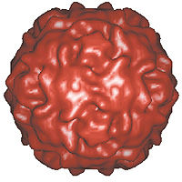
The first images of a polio virus as it infects a human cell have been captured by researchers at Harvard Medical School.
The paralyzing disease has been eradicated from Western countries with vaccines, but biologists still want to know how it gets into the cells of the intestines, from where it makes its way to the nervous system. They believe that other viruses use similar break-in methods to cause different maladies, including encephalitis, paralysis, diabetes, and heart ailments.
” Understanding these viruses gives you a route to potentially making drugs to thwart them,” notes James Hogle, Harkness Professor of Biological Chemistry and Molecular Pharmacology. Hogle and colleagues at Harvard and the National Institutes of Health made the three-dimensional images by crystallizing the virus, irradiating it with x-rays, and then fitting the x-rays together under an extremely powerful microscope.
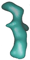
The result shows that the virus throws out minute protein threads that embed themselves into the thin, soft envelope surrounding the cell. Besides securely gripping the cell, this anchorage may create tiny pores for viral genes to enter and start reproducing more viruses.
The polio virus boasts a surface wrinkled with minute canyons. One of many receptors that fit into the canyons and hold the virus securely to the surface of the cell it infects.