

Photos by Scott Chimileski
Health
Life of the party
Microbial palettes and patterns make strange, spectacular slideshow
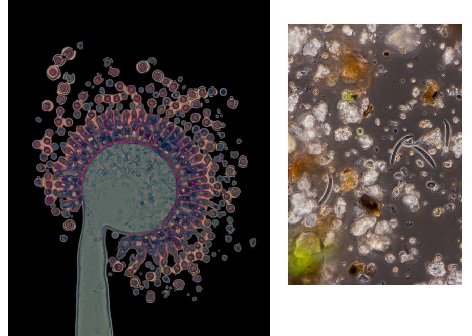
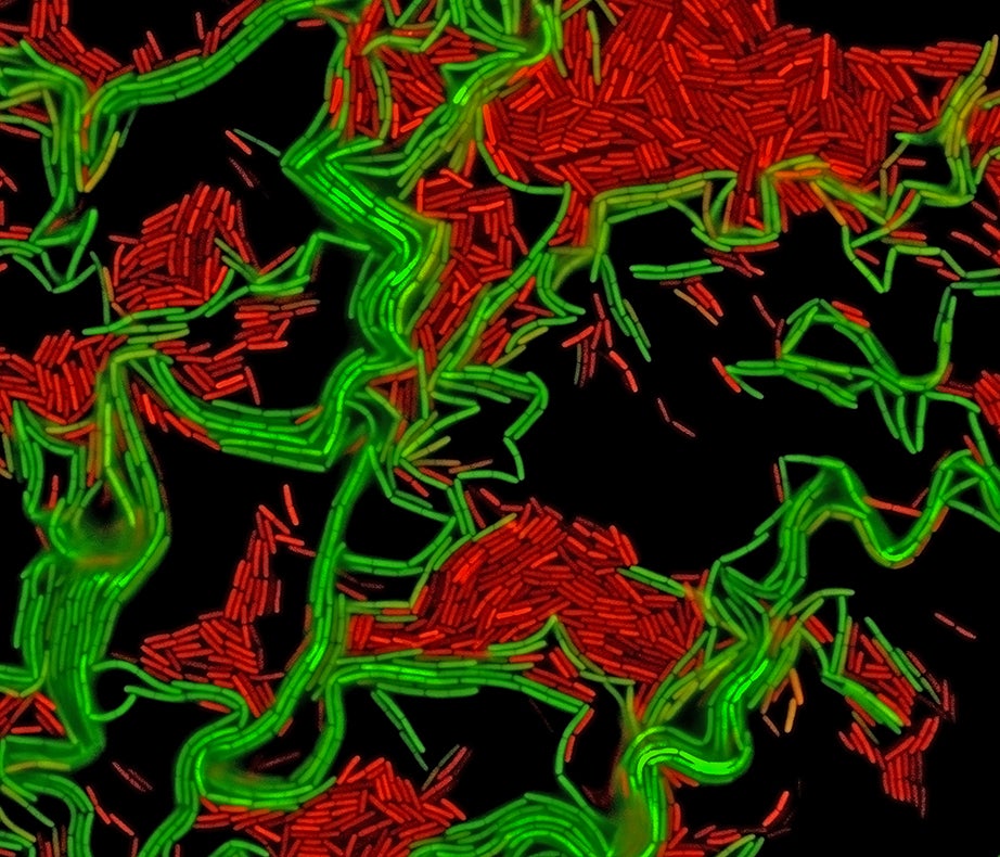
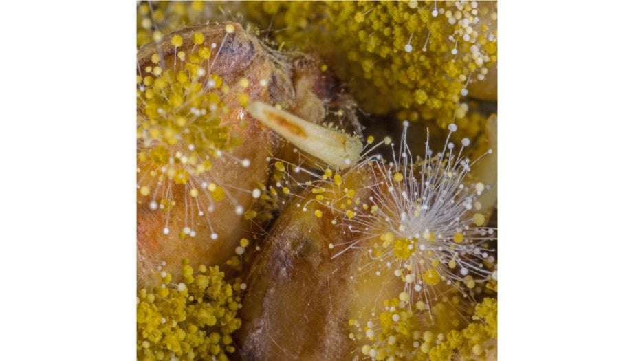
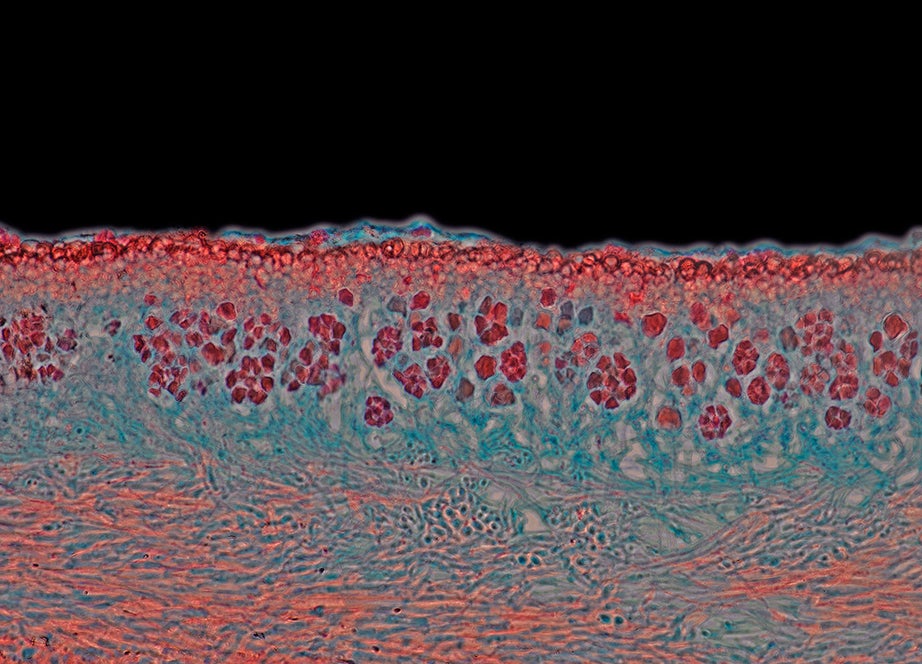
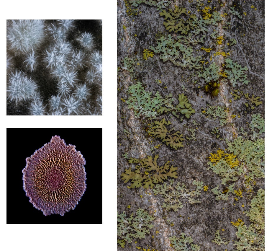
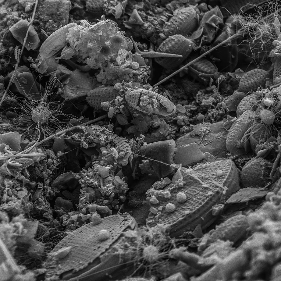
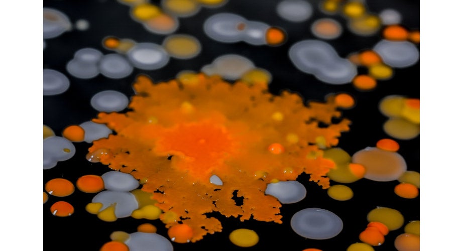
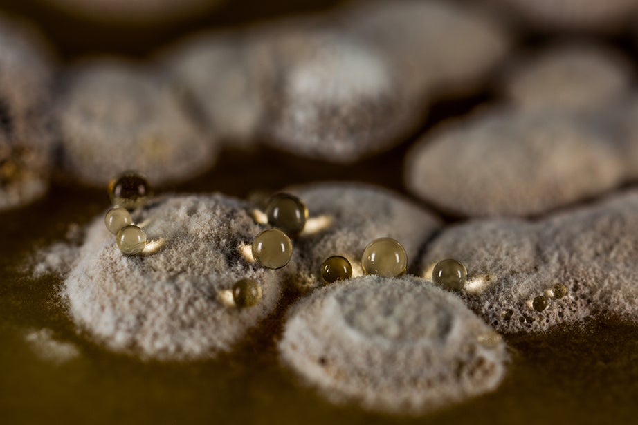
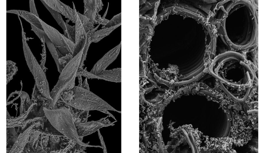
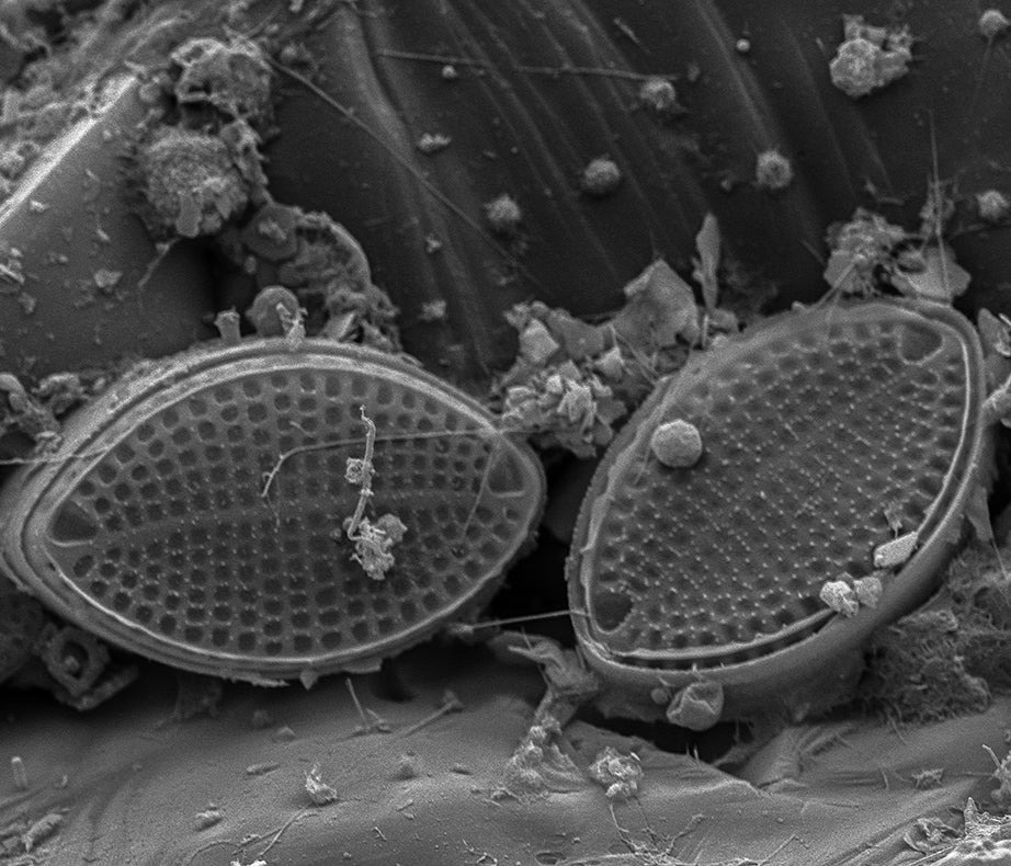
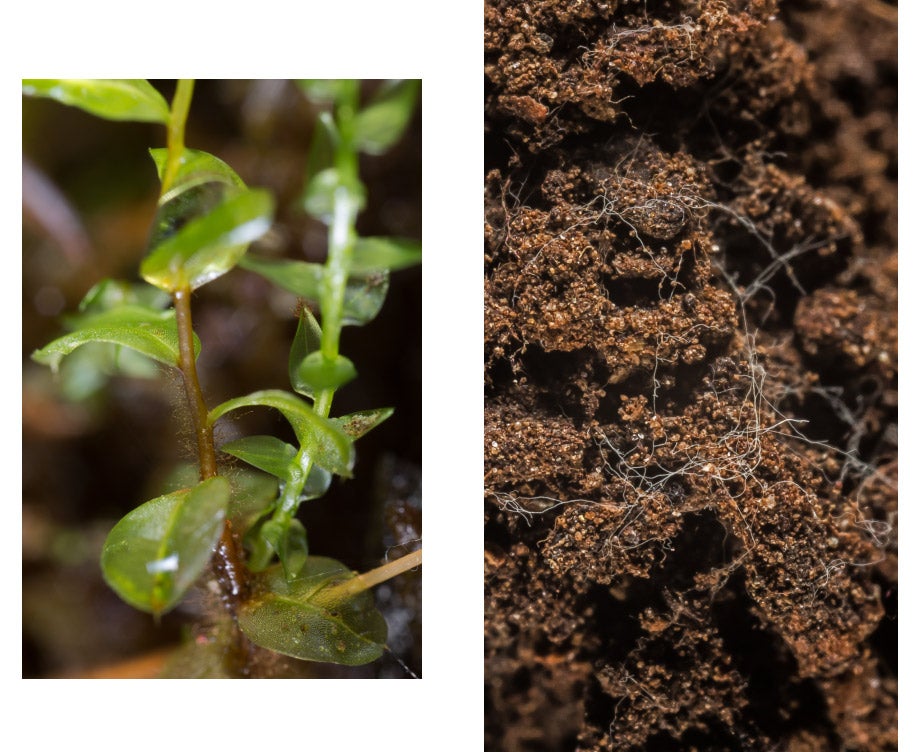
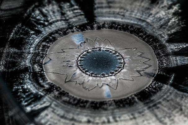
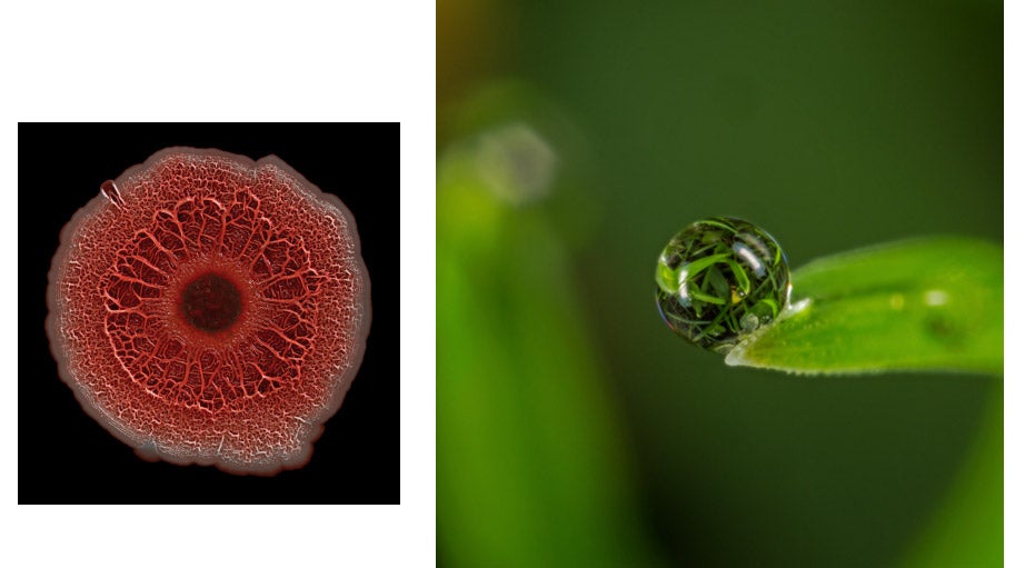
A hidden world of vibrant colors, dynamic movements, and extraordinary shapes has come alive at Harvard.
Researchers Roberto Kolter and Scott Chimileski’s large-scale photographs illustrating the intricate social and multidimensional wonder of the small-scale world of microbes are on display in the Science Center show “Scale: A Matter of Perspective” through Dec. 9. The stunning images, which blend art and science, also comprise the exhibit “World in a Drop: Photographic Explorations of Microbial Life” at the Harvard Museum of Natural History through Jan. 7.
For a hands-on experience, educators, researchers, and the Art+Bio Collaborative will hold a Microbes Mini-Festival on Sunday from 1 to 4 p.m. Visitors can observe thriving microbial colonies on cheese rinds, and watch the aquatic micro-animals known as “waterbears” prowl a landscape the unaided human eye can’t see. The opportunity to work on a collaborative art project detailing a large microbial community and interact with fungi is part of the fun.
The images below give a glimpse of what keeps our ecosystems and our lives in balance. These stunning pictures invite people to explore the significant, structural world of the unseen.
— Deborah Blackwell
Share this article